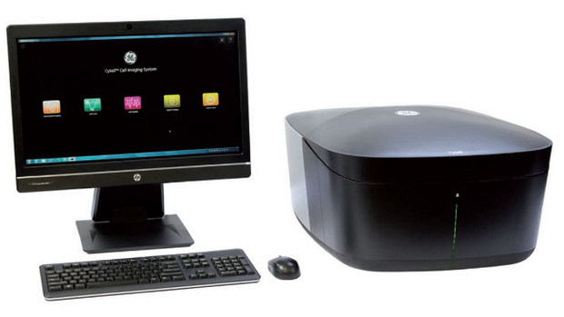
Cytell Cell Imaging System
GE Healthcare launches Cytell Cell Imaging System for compact, app-driven, automated imaging - Streamlines data acquisition, analysis and visualisation
Cytell Cell Imaging System is an intuitive benchtop instrument equipped with on-board data analysis and visualisation tools. It streamlines and simplifies routine assays such as cell cycle and cell viability and efficiently delivers high quality, quantitative results.
Key to the simplification are the on-board Bioapps; pre-configured, easy to use software modules that automate all the steps from imaging and data acquisition through data analysis, visualisation and report generation. The Cytell cell imaging system is compact and small enough to sit on a benchtop.
Cytell can image a wide variety of sample formats including slides, petri dishes, T-flasks and microplates. Compatible GE Healthcare Life Sciences reagents and kits are available, although Cytell is an open system and therefore provides the flexibility to create customized protocols based on other suppliers’ reagents.
BioApps currently available include:
- Digital Imaging BioApp: Acquire fluorescence and transmitted light images of samples in plates, flasks, Petri dishes, or slides.
- Automated Imaging BioApp: Automatically acquire high-quality fluorescence and transmitted light images from an entire multiwell plate in minutes.
- Quick Count BioApp: Obtain accurate cell counts and viability and concentration estimations based on fluorescence intensity using a disposable hemocytometer.
- Cell Cycle BioApp: Analyze cell cycle phase distribution in multi-well format using a DNA-binding fluorophore.
- Cell Viability BioApp: Run two- or three-color assays in multiwell formats with subpopulation gating to determine the percentages of live, dead, and non-viable cells.
For further information please visit http://www.gelifesciences.com/Cytell


 Unsplash+
