Generation of Functional Monoclonal Antibodies by Single B Cell Cloning
B cells play a key role in the immune system given their primary role in antibody production. Antibodies, also known as immunoglobulins, are glycosylated protein molecules present on the surface of B cells serving as antigen receptors, or are secreted into the extracellular space where they can bind and neutralize their target antigens. B-cell development begins in the bone marrow and progresses sequentially through pro-B, pre-B and immature B cell stages with the expression of surface IgM, a mature B-cell receptor (BCR). Each B cell produces a single species of antibody, each with a unique antigen-binding site. When an antigen binds to the B-cell surface it initiates B-cell maturity and division into an antibody-secreting effector cells, called plasma cells, which secrete millions of antibodies into both the bloodstream and lymphatic system (Figure 1).
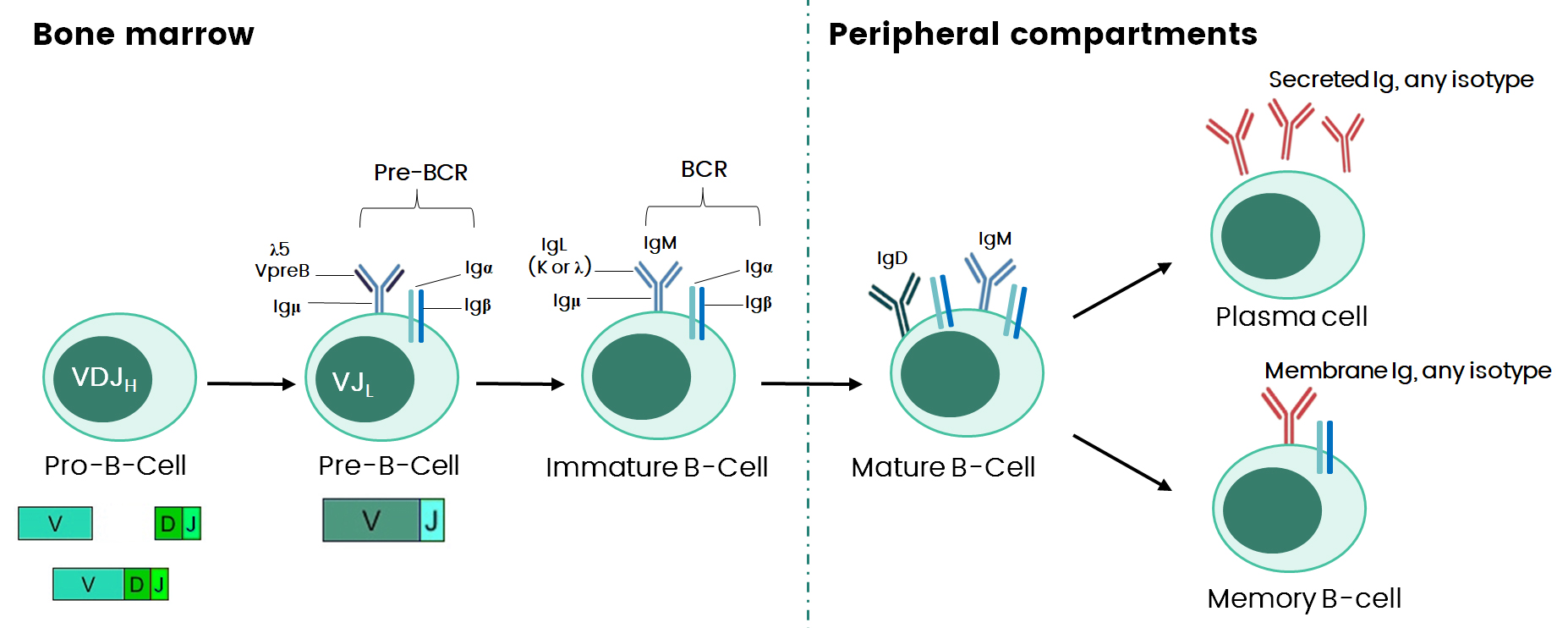
Figure 1. The Main Stages of B-cell Development.
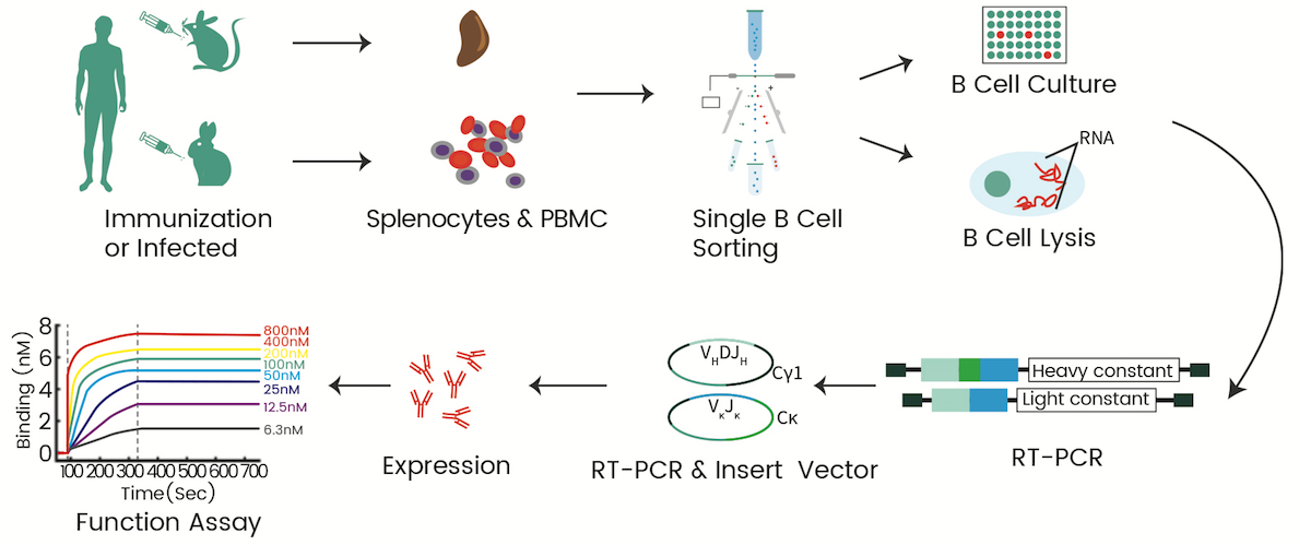
Sino Biological leverages a single B-cell sorting platform to provide single B-cell antibody production services involving single B cell isolation, culture, sequencing, cloning and screening of select single B-cell antibodies (Figure2)
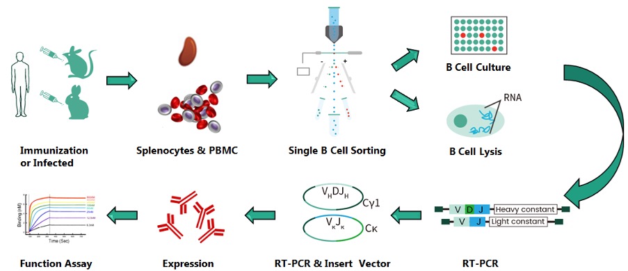
Figure 2. Workflow of Sino Biological’s Single B-Cell Antibody Discovery Platform.
Single B-cell isolation can be performed by selection of antigen-specific B-cells using antigen-coated magnetic beads and fluorochrome-labeled antigens via multi-parameter fluorescence-activated cell sorting (FACS), the hemolytic plaque assay, and fluorescent foci methodology. With FACS technology, cells can be sorted and clearly distinguished based upon the expression patterns of specific cell surface markers. In general, B-cells at any stage can be sorted, but class-switched memory B cells and antibody-secreting cells (ASCs, i.e. plasmablasts and plasma cells) are of special interest in obtaining relevant monoclonal antibodies (mAbs) as they bear somatically mutated B-cell antigen receptors (BCRs) exhibiting considerably high affinities.
Memory B-cells can be polyclonally stimulated in vitro to induce secretion of detectable amounts of antibody. Firstly, stimulation can be induced by a mixture of the agonist and recombinant cytokines for several days. Subsequently, supernatants of the microwells are screened by ELISA for the presence of antibodies of the desired specificity followed by lysis of B-cells from selected wells.
Single-cell cDNA synthesis is usually performed in 96-well plates with full-length Ig gene transcripts amplified by nested RT-PCR. Typically, forward primer mixes complementary to the corresponding IgH and IgL V gene leader sequences and a single reverse primer specific to the constant region sequence are employed for nested RT-PCR. If necessary, for example, when isotype independent cell sorting is performed, mixed reverse primers can be used for the amplification of IgH chains with different constant regions. In the second round, nested primers or primer mixes are used to increase sensitivity and specificity. During this step, homologous armsfor subsequent cloning steps can be incorporated into the amplicons. Linear expression cassettes can subsequently be directly transfected into mammalian cells for efficient in vitro expression of monoclonal antibodies.
To determine the reactivity profile and biophysical characteristics of antibodies, target proteins have to be expressed, purified and antibodies tested on these targets in various assays. The most common choices for expression systems are transient and/or stable mammalian cell systems (e.g. HEK 293, CHO cells). In mammalian cells, antibody expression can exist in complete IgG format.
The COVID-19 pandemic caused by SARS-CoV-2 has influenced both the health care systems and economies around the world, and has posed challenges to vaccines, drugs and diagnostics development. Since the outbreak, scientists at Sino Biological have been actively tracking the change and movement of the virus to develop potent anti-spike neutralizing antibodies from immunized animals.
This study was designed to identify anti-SARS-CoV-2 neutralizing antibodies from a rabbit immunized with the recombinant receptor binding domain (RBD) domain of COVID-19. Neutralizing antibodies were discovered by high-throughput screening of antigen-specific B-cells from rabbit primordial bone marrow cells (PBMC)s. Single-cell RT-PCR combined with fluorescence-activated cell sorting (FACS) and B-cells activated in vitro allowed for the determination of antibody sequences in roughly two weeks. This was accomplished by performing nested PCR on single antigen-binding memory B cells after single-cell sorting and cell culture. Fifty-four antigen-binding B cell clones expressing antigen-specific antibodies were identified from 600 single B-cell supernatants. The purified monoclonal antibodies were tested for SARS-CoV-2 RBD/spike reactivity by ELISA and bio-Layer interferometry (BLI), and 27 RBD-binding antibodies were identified.
All ELISA-positive anti-SARS-CoV-2 mAbs were further screened for neutralizing ability using a SARS-CoV-2 pseudovirus system, which was produced by co-transfection of HEK-293 T cells with plasmids encoding lentiviral packaging and luciferase reporters. Four of the ELISA-positive anti-SARS-CoV-2 antibodies showed potent neutralization ability with an IC 50 lower than 0.15μg/mL against SARS-CoV-2 pseudovirus (Figure 3). All four anti-SARS-CoV-2 antibodies bound strongly to the RBD with close to pM KD, revealed by BLI (Figure 4).
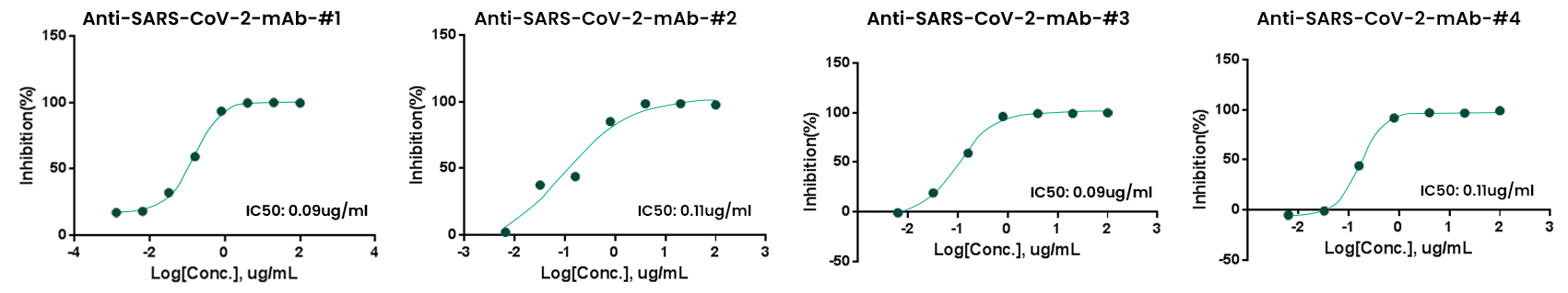
Figure 3. Anti-SARS-CoV-2 Monoclonal Antibody Neutralization with Pseudovirus.
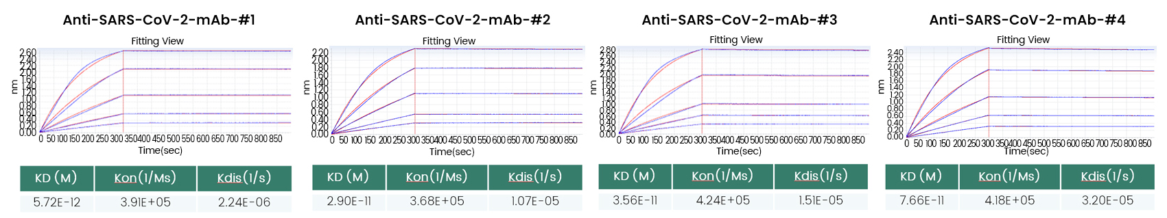
Figure 4. Anti-SARS-CoV-2 Monoclonal Antibodies Affinity as Assessed by BLI.
T-cell immunoglobulin and mucin domain-3 (TIM-3), an important immune checkpoint, has been shown to inhibit cancer immunity. TIM-3 is primarily expressed on immune cells, particularly on dysfunctional and exhausted T-cells, and engagement of TIM3 with its ligands promotes TIM-3-mediated T-cell inhibition. Antagonistic ligand-blocking anti-TIM-3 antibodies have the potential to abrogate T-cell inhibition, activate antigen-specific T-cells and enhance anti-tumor immunity.
Sino Biological has developed anti-TIM-3 monoclonal antibodies that were derived from rabbits immunized with the His-tagged recombinant human TIM-3 extracellular domain (ECD) protein. Following B-cell sorting and proliferation, 200 antigen-specific antibodies were identified from 1100 single B-cell supernatants. The kinetics and affinity of 63 purified ELISA-positive anti-TIM-3 monoclonal antibodies were evaluated by bio-Layer interferometry (BLI) technology. Among the ELISA-positive antibodies, five of them showed the best affinity with a range between 10-11 and 10-12M (Figure 5).
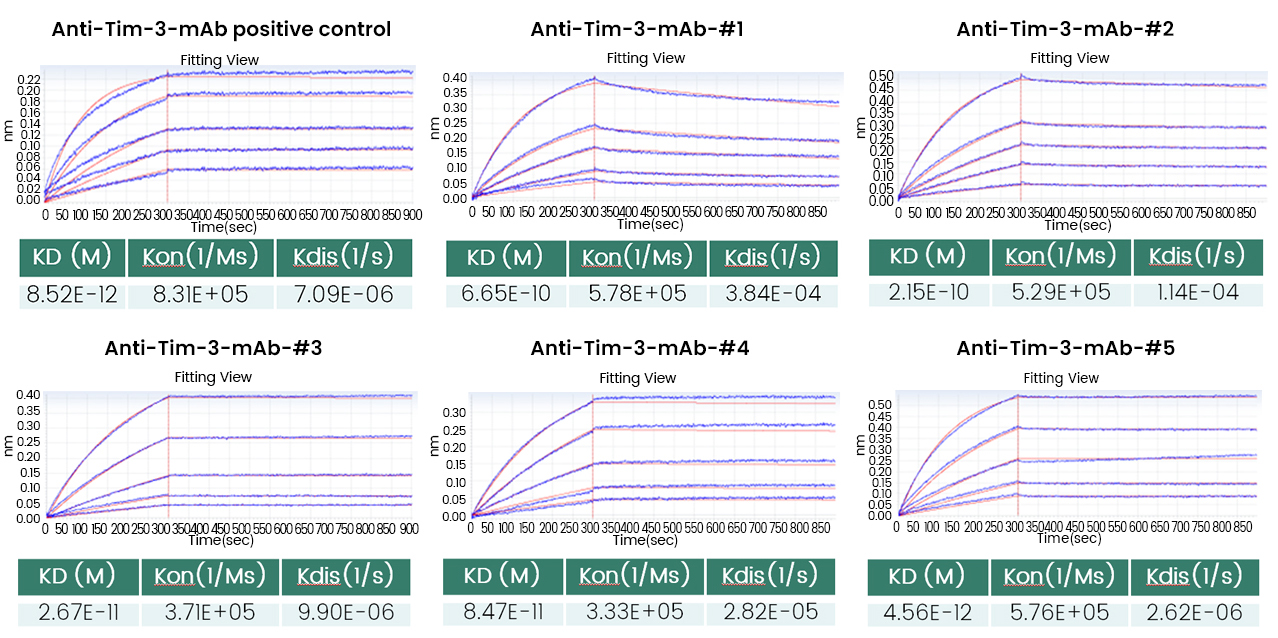
Figure 5. Anti-Tim-3 Monoclonal Antibody Affinity as Assessed with BLI.
To evaluate the ability of the five anti-TIM-3 monoclonal antibodies to block the interaction of TIM-3 with its ligand phosphatidylserine (PtdSer), a blocking assay was performed using a bioluminescent reporter cell-based system. Five anti-TIM-3 mAbs was able to efficiently block the interaction between TIM-3 and PtdSer in a dose-dependent manner, with the IC50 of 0.070 ± 0.010 μg/mL, close to that of positive control (Figure 6).
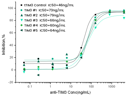
Figure 6. Anti-Tim-3 Monoclonal Antibody Ligand-blocking Cell-based Assay.