Google maps for tumours and organs
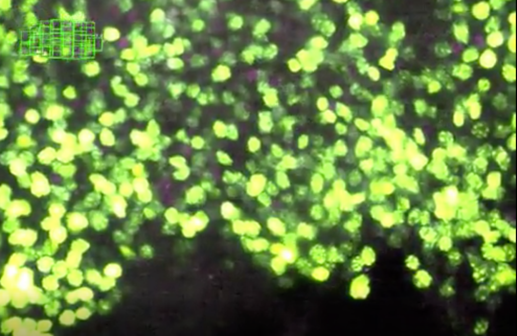
Theoretically, light sheet microscopy can be used i.e. to observe in 3D, which parts of a mouse brain or a whole tumour are penetrated or affected by a drug by making a tissue or tumour transparent first by a special sample preparation technique and scanning sheet by sheet of the tissue afterwards, reconstructing a 3D image from the terabytes of data in the end. So far, management of the huge amount of data made it nearly impossible to navigate through the 3D reconstruction.
A team headed by Stephan Preibisch at Berlin-based Max-Delbrück Center for Molecular Medicine now report in Nature Methods that it has developed a software tool that allows managing the terabytes of image data. "The problem, however, was that the procedure generates such large quantities of data – several terabytes – that researchers often struggle to sift through and organize the data," says Preibisch.
To create order in the chaos, Preibisch’s team developed a software program that after complex reconstructing the data resembles somewhat Google Maps in 3D mode. "You can not only get an overview of the big picture, but can also zoom in to specifically examine individual structures at the desired resolution," explains Preibisch, who has christened the software "BigStitcher."
The researchers show in their paper that algorithms can be used to reconstruct and scale the data acquired by light-sheet microscopy in such a way that renders a supercomputer unnecessary. "Our software runs on any standard computer," says Preibisch. "This allows the data to be easily shared across research teams."
The development of BigStitcher took ten years. Today, BigStitcher can not only visualize on screen the previously imaged samples in any level of detail desired. "The software automatically assesses the quality of the acquired data," says Preibisch. This is usually better in some parts of the object being studied than in others. "Sometimes, for example, clearing doesn’t work so well in particular area, meaning that fewer details are captured there," he adds.
"The brighter a particular region of, say, a mouse brain or a human organ is displayed on screen, the higher the validity and reliability of the acquired data," says Preibisch. And because even the best clearing techniques never achieve 100 percent transparency of the sample, the software lets users rotate and turn the image captured by the microscope in any direction on screen. It is thus possible to view the sample from any angle.


 Bayer AG
 Picture from Ferdinand Stöhr on Unsplash