A modern electron microscope (cryo-EM) available for industry at the Solaris Center in Krakow, Poland
Easier access for central European companies to cryo-EM technique! A modern Thermo Scientific Glacios transmission electron microscope has just been installed at Synchrotron Solaris in Poland and it is open for industrial users.
Cryo-EM – a revolutionary technique
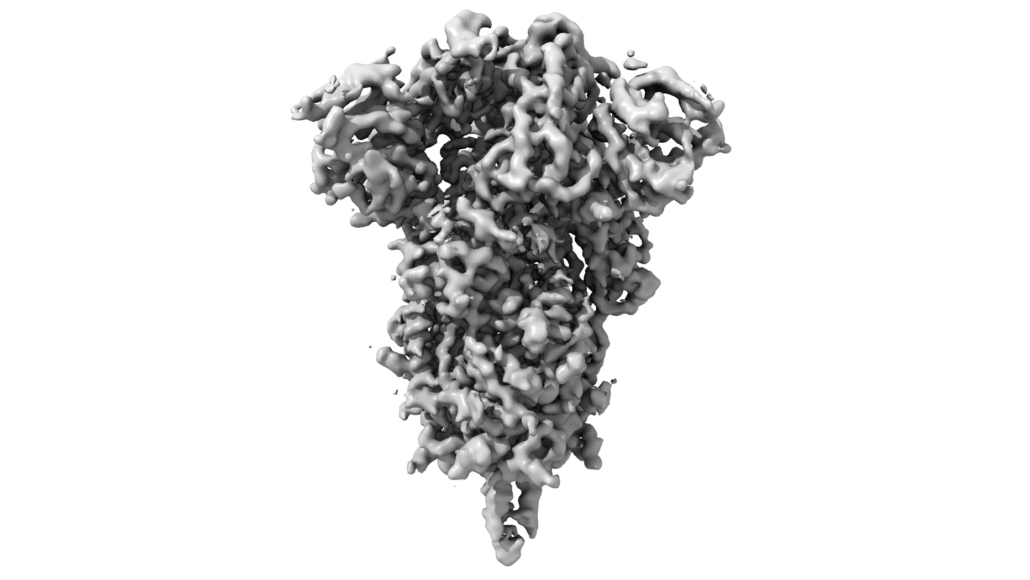
In recent years, the technique of cryogenic electron microscopy (cryo-EM) has revolutionized research in the field of structural biology. The 2017 Nobel Prize in Chemistry confirmed that this technique bring undisputed changes to structure determination process. The development of this method has allowed to solve the structures of proteins or protein complexes, which are crucial for the world of science and medicine, such as the ribosome or membrane proteins. Until now, such results were unattainable by crystallography methods. A small amount of protein is needed to prepare the grid, which is frozen in a very thin, amorphous layer of the solution. This way the proteins do not have to take a crystalline form. This enables the imaging of the structure in the native state, which is closest to the natural one, thus giving this method an advantage over crystallography.
Research using the cryo-EM technique allows for a deep insight into the interaction of protein complexes with other proteins, nucleic acids, antibodies and the study of protein-inhibitor or protein-active molecule interactions. As a result, the technique is of particular interest to drug design companies.
Cryo-EM for small business and industry
Responding to the great interest in technology among biotechnological, pharmaceutical and cosmetic companies, Solaris Center in Krakow started to provide commercial services using on the newly purchased Thermo Scientific Glacios microscope. Thus, European entrepreneurs gained the opportunity to use this modern technique in Poland.
The Glacios microscope, with an accelerating voltage of 200 kV, allows for a reconstruction with a resolution of 2 Å. The microscope is equipped with two cameras – 16-bit, CMOS type, adapted to conduct electron microdiffraction studies (Thermo Scientific CetaD), with a resolution of 14 MP and a camera for direct electron detection, with a resolution of 14 MP (Thermo Scientific Falcon 4). In addition, the microscope’s equipment allows for tomography mode (cryo-ET) and the use of modern microcrystal electron diffraction imaging techniques (microED).
The Solaris Center houses not only the largest and most modern research infrastructure in Poland, but more importantly also a team of world-class professionals. Some of the data has already been published in reputable scientific journals such as Nature Communications, Journal of Virology or Science Advances. The extensive experience of Solaris specialists includes not only research using on innovative Thermo Scientific Krios and Glacios microscopes, but also working with a variety of research samples – from artificial protein constructs in the form of nanocages, to proteins of viral origin.
Fast and effective solving of protein structures
The Solaris Center offers two standard service packages. The first one allows the characterization of a sample and the optimization of experimental parameters by preparing and measuring 6 grids with different protein concentrations. This allows to determine the best measurement conditions and to initially collect data for the assessment of the reconstructive potential. The second packet includes a complete reconstruction of the structure on the basis of the best selected grid from screening. The center offers not only the possibility of measurements, but also the preparation of grids, freezing the samples, storage, reconstruction and solving the structure, but also consultation of the obtained results.
We encourage you to contact with our specialist –
dr Piotr Ciocho?
+48 12 664 41 11
e-mail: industry.solaris@uj.edu.pl
More info –
https://synchrotron.uj.edu.pl/przemysl/dostepne-uslugi/cryoem
The project for the development of the scientific and research infrastructure of the Jagiellonian University by creating a cryo-electron microscopy laboratory, enabling measurements used in business activities, is co-financed by the European Union – the European Regional Development Fund under Measure 4.2 of Smart Growth Operational Programme 2014-2020.


