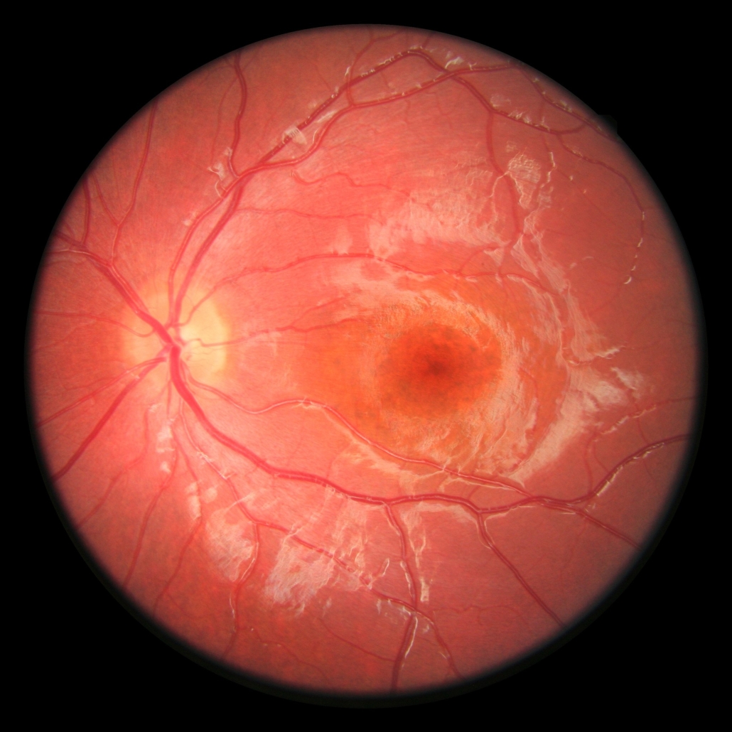
Sheets of eye cells improve retina repair
Scientists have generated sheets of human eye cells arranged on a biological scaffold, which they used to successfully treat vision disorders in rats.
According to the team led by French researcher Olivier Goureau from INSERM Paris, tissue-engineered cell sheets may improve the efficacy of cell therapy for tackling retinal degenerative diseases. Based on promising preclinical findings, the researchers are scaling up their protocol with the goal of launching a phase I/IIa clinical trial to prevent vision loss for patients with retinitis pigmentosa – a genetic disorder that leads to progressive deterioration of light-sensing cells in the eye.
Regenerative medicine strategies to replace dead or defective eye cells have emerged as promising options for treating retinitis pigmentosa as well as other debilitating vision disorders like age-related macular degeneration, which affects millions of people worldwide. Although injections of stem cell-derived eye cells have proven safe in clinical trials, so far the transplants rapidly die off, offering limited potential benefit.
Reasoning that a three-dimensional structure might help transplants perform better, first author Karim Ben M’Barek and colleagues engineered supportive scaffolds composed of stem cell-derived eye cells overlaid onto sheets of human amniotic membrane and gelatin. At all stages, the researchers ensured that the grafts were free of contaminants or potentially tumour-forming cells. In rat models of RP, transplants of the new sheet-grafts led to improved vision and light responsiveness compared to animals that received injections of freely-suspended eye cells. The researchers note that tissue banks of human amniotic membrane are widely available, and that their eye cell production protocol was compliant with good manufacturing practices, paving the way for clinical applications.


 ©FabienMalot
 Lonza Group
 Vetter Pharma