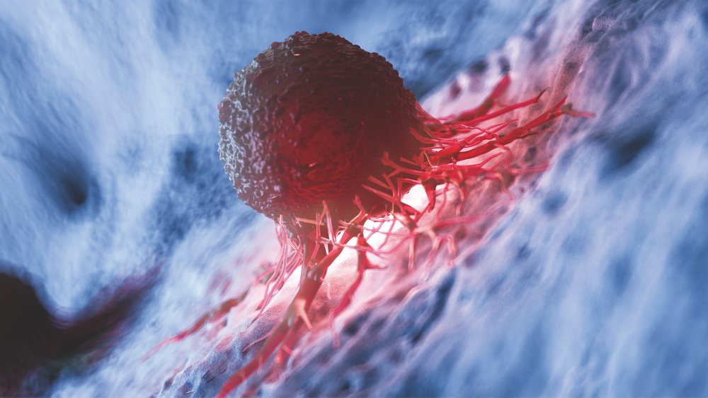
A Holy Grail in the quest to end cancer
When he went in for a routine check-up, Peter Widener discovered he had pancreatic cancer. It’s one of the worst diagnoses a doctor can hand out. At Peter’s stage of the disease, most people are dead within a year. – but he didn’t have any symptoms. In fact, the 75-year old retired architect still enjoys sports and meeting new people. So he was glad when his oncologist said he didn’t want to conduct a “10-hour surgery that is going to make you feel like you’ve already died” as long as the “tumour is as inactive as it seems to be at the moment.”
Peter was luckier than most patients with his diagnosis, because their primary tumour generally spreads early and returns – in a much more malignant form – at distant sites. In breast cancer, in around a third of the women with the still apparently localised form of the disease, cancer cells have already spread to distant sites by the time they are diagnosed. Many women relapse 12–18 months after surgery.
While cancer survival rates overall have dramatically improved thanks to better diagnoses and drugs that stop tumour proliferation, survival rates for people with metastases have improved little. About 50 years after US President Richard Nixon kicked off the ‘War on Cancer’ by signing the National Cancer Act of 1971, metastasised bone, brain, lung or liver cancer still leads to over 90% of all cancer deaths, stressed researchers from the German Cancer Research Centre (DKFZ) at a workshop on metastasis in late January. While there’s no drug on the market that can prevent it, a small number of oncologists have left mainstream research – which mainly targets primary tumours – and turned to unravelling the mechanisms behind metastatic dormancy, metastasis, and its triggers, adding a couple of preclinical candidates to pipelines along the way.
When it comes to metastatic cancer, most drugs that are prescribed target primary tumours instead of metastasis formation. The only real market currently out there is not for causative therapies, but for symptom treatments – including medications aimed at pain control, preventing fractures or improving functional disabilities. Inkwood Research projects that market will continue to grow between 2018–2026 at a CAGR of 7.13%. As metastasis is currently the leading cause of death for the 14 million patients newly diagnosed with cancer annually, the focus is now turning to approaches that target metastases directly. Most of the work being done in the field, however, is still very much in the discovery stage.
Signals that guide metastasis
Among the pioneers seeking to understand the roots of metastasis is the head of the Ludwig Center at MIT’s Whitehead Institute. Robert Weinberg discovered the first oncogene, and co-authored Cell’s most-cited paper ‘Hallmarks of Cancer’. “We are just beginning to understand which signals trigger metastasis,” he cautions. In a mouse model that mimics metastatic dormancy, his team has for the very first time established a causal link between post-surgical wound healing, inflammation, and metastasis – providing a glimmer of hope to patients apprehensive their cancer might come back. In the mouse model (Sci. Transl. Med., doi: 10.1126/scitranslmed.aan3464), Weinberg’s team demonstrated how the systemic inflammatory wound healing response following breast cancer surgery triggered the outgrowth of tumour cells at distant anatomical sites. According to Jordan Krall, the paper’s first author, surgical wounds specifically induced an increase in the number of circulating neutrophils and pro-inflammatory monocytes, and boosted levels of interleukin-6, CCL2 and G-CSF. Such signals are believed to prepare a niche for metastatic precursors, helping them home in on distant sites. In mice, the researchers have now succeeded in preventing the process. If Weinberg and Krall administered their model up to four days post-surgery with meloxicam – a nonsteroidal anti-inflammatory drug (NSAID) – mice showed significantly reduced numbers of metastases. This suggests that one day, a cheap and common drug may improve breast cancer outcomes. Andreas Trumpp adds that “in 2008, we showed that the most potent (normal) stem cells are in dormancy, protecting themselves from mutations, but can be reactivated by inflammatory processes.” The Managing Director of the Heidelberg Institute for Stem Cell Technology and Experimental Medicine (HI-STEM) says that “during reactivation, DNA damage increases due to rapid (SOS) entry into genome replication.”
Pursuing the fundamentals
In mid-February, Hibercell Inc – the first company dedicated to detecting and targeting dormant cancer cells – took off with a US$60.8m Series A round of financing to push three preclinical programmes in solid cancer relapse and one in blood cancer metastasis. Spun out from the lab of Julio Aguirre-Ghiso
(Icahn School of Medicine at Mount Sinai), Hibercell has identified a “dormancy signature” enriched in dormant tumour cells (DTCs) from patients asymptomatic for up to 18 years. This signature can act as a predictor for prolonged metastasis-free periods in different cancers. To form distant metastases, cells from the primary tumour need to migrate into the bloodstream, disseminate, and then penetrate their preferred target tissue. However, only a small fraction of these circulating tumour cells (CTCs) released from cancers of epithelial origin seem to be able to form mestasta-
tic lesions. Most seem to die in the bloodstream. Several research groups have been hunting for the pool of metastasis-initiating cells (MICs) to learn more about them. Understanding the molecular features of this population of stem-cell-like CTCs could prove to be a cornerstone in developing metastasis-tailored therapies.
Preventing blood-borne metastases
In 2013, Trumpp and his colleague at HI-STEM Irene Baccelli identified CTCs in breast cancer patients that were able to initate metastasis in vivo (Nature Biotech. 31 (2013), p. 539–544). Two years later, scientists at Massachusetts General Hospital in Boston reported the isolation of oligoclonal clusters of 2-50 CTCs (CTC clusters) from breast cancer patients with a microfluidic device. In their patent, medical researchers Nicola Aceto, Daniel Arie Haber and Shyamala Maheswaran wrote that “CTC-clusters-enriched patients progressed to metastasis more rapidly (mean progression-free survival (PFS): 76.1 days) than those enriched with single CTCs (mean PFS: 160.6 days).“ Their results indicate that the presence of CTC-clusters is associated with faster metastatic spread and disease progression in breast cancer patients. Further analyses revealed that CTC-clusters appear to originate from regions of the primary tumour characterised by the high expression of plakoglobin, a cell-to-cell junction mediator. In animal models, plakoglobin knockdown in the primary tumour decreased the number of tumour-derived CTC-clusters, as well as lung metastases.
At the end of January, Aceto – who has since moved to the University of Basel – published novel details on CTC clusters. His team reported that formation of CTC clusters vs. single CTCs was associated with hypomethylation of binding sites for factors promoting the stemness (dedifferentiation and proliferation) of circulating tumour cells. A list of these factors includes OCT4, SOX2, NANOG and the transcription regulator SIN3A. In breast cancer patients, hypomethylation of these factors significantly decreased progressionfree survival.
In a screen of 2,468 FDA-approved compounds, Aceto’s group identified six compounds that reduced the size of CTC clusters. Na+K+ ATPase inhibitors ouabain and digitoxin were most effective. As intracellular Ca2+ levels increase with rising concentrations of the Na+K+ ATPase inhibitors, Aceto and colleagues hypothesise that increased Ca2+ triggers the loss of cell-cell junctions between cancer cells. CTC dissociation also led to epigenetic remodelling of the previously hypomethylated transcription factor binding sites. In preclinical models, daily ouabain administration over three weeks reduced the abundance of CTC clusters, increased the fraction of single CTCs and was accompanied by a 80.7-fold reduction of the total metastatic burden – a tremendous reduction of metastasis seeding. Aceto says he next wants to confirm his findings and check compound safety in patients.
Analysing MIC’s vulnerabilities
Other European groups, like that headed up by Trumpp or the one led by Salvator Benitah at IRB Barcelona have provided evidence that even single metastasis-initiating cells (MICs) can trigger metastasis development. Trumpp thinks CTC clusters might serve to block differentiation of CTCs, or possibly protect them from immune attacks in blood. His group has established apheresis methods to isolate large amounts of rare CTCs from the blood of relatively stable patients with metastasising cancer, and to culture them in order to carry out single-cell transcriptomics or other biochemical characterisations of hidden MICs in this cell population. According to the DKFZ, a first application of this liquid biopsy platform will track molecular alterations in CTCs from breast cancer patients. The goals are to select optimal individual treatment options, and to set up a comprehensive biobank with biospecimens and clinical results for comparative analyses.
Benitah’s group most recently described a subpopulation of MICs: non-mesenchymal CD44bright cells in human oral carcinomas that express high levels of the fatty acid receptor CD36 and lipid metabolism genes. While palmitic acid or a high-fat diet specifically boosted the metastatic potential of CD36-positive MICs, neutralising antibodies that blocked CD36 caused a nearly complete inhibition of metastasis in immunodeficient or immunocompetent orthotopic mouse models of human oral cancer – without any side effects. Clinically, the presence of CD36-positive MICs correlates with a poor prognosis for numerous types of carcinomas, and inhibition of CD36 impairs metastasis, at least in human melanoma- and breast-cancer-derived tumours.
Locking the endothelium
Another strategy for fighting metastasis at its earliest stages is to prevent MICs or CTC clusters in the blood from homing in on distant sites. According to Andreas Fischer from the DKFZ, the primary tumour sends out messengers, exosomes or inflammatory mediators to prepare the groundwork for pioneering cancer cells. “Some organs respond to it by remodelling, dampening the immune response, angiogenesis or the formation of docking sites in blood vessels,” he says. “This facilitates the invasion and survival of tumour cells in these organs.” Two years ago, Fischer’s team reported that enhanced NOTCH1 signalling in the primary tumour and in the endothelium of blood vessels facilitated metastasis. NOTCH1 signaling weakened endothelial cell junctions and boosted VCAM1 expression, which helps CTCs to attach to the layer. Both factors eased migration of tumour cells and immune-dampening across the vessel wall into lung tissue. However, when Fischer and colleagues administered antiNOTCH1 or anti-VCAM-1 antibodies to mice, the number of lung metastases the animals developed decreased. However, “NOTCH1 is a universal signal molecule, and thus can’t be used over extended periods of time, because it triggers congestive heart failure,“ Fischer stresses. He told European Biotechnology that nOTcH also has other pleiotropic effects such as “preventing excessive angiogenesis leading to the formation of dysfunctional blood vessels, and maintaining the structure and function of liver sinusoidal endothelial cells.”
One speculation is that drug candidates able to restore the endothelial barrier function may also be able to reverse NOTCH1-triggered endothelial dysfunction. Currently, an antibody (adrecizumab) that restores the barrier function by concentrating the vasoactive peptide hormone adrenomedullin (ADM) in the vessel lumen is in Phase II efficacy testing to prevent the life-threatening drop in blood pressure, and thus improve overall survival in patients with septic shock.
Stopping dedifferentiation
Another strategy to eliminate MICs has been developed by Gerhard Christofori at the University of Basel (Cancer Cell, doi: 10.1016/j.ccell.2018.12.002). In February, his team reported they were able to stop the dedifferentiation process leading to epithelial-mesenchymal transition (EMT) in breast cancer cells. After a thorough analysis of underlying MIC transcription patterns, his team used the MEK inhibitor trametinib and the antidiabetic rosiglitazone to coax human breast-cancer cells to turn into fat cells in mice that bore human tumours. Treatment blocked the invasion of tumour cells into tissue and the formation of metastases. Although not every cancer cell in the study changed into an adipocyte, the ones that underwent adipogenesis didn’t change back. “The breast cancer cells that underwent an EMT not only differentiated into fat cells, but also completely stopped proliferating,” stressed Christofori. What’s exciting about the work is that the two drugs involved have already been FDA-approved, so it should be easier to get them into clinical trials in people. “In the future, this innovative therapeutic approach could be used in combination with conventional chemotherapy to suppress both primary tumour growth and the formation of deadly metastases,” Christofori concluded.
Progress in CTC isolation techniques has enabled a lot of studies in blood-borne metastasis. When spreading from primary tumours, however, cancer cells also follow other routes, including:
- Transcoelomic spread of malignant cells into body cavities can occur when they penetrate the surface of the peritoneal, pleural, pericardial or subarachnoid spaces.
- Lymphatic spread allows the transport of tumour cells to regional lymph nodes near the primary tumour, and ultimately to other parts of the body. Lymphatic spread is the most common route of initial metastasis for carcinomas.
- Haematogenous spread is the typical route of metastasis for sarcomas, but it is also the favoured route for certain types of carcinomas like renal cell carcinoma. Because of their thinner walls, veins are more frequently invaded than arteries, and metastasis tends to follow the pattern of venous flow.
- Canalicular spread: Some carcinomas may metastasise along anatomical canalicular spaces. These spaces include for example the bile ducts, the urinary system, the airways and the subarachnoid space. However, often it remains unclear whether simultaneously diagnosed tumours of a canalicular system are one metastatic process or in fact independent tumours caused by the same agent
Cancer of unknown primary origin (CUP)
In theory, metastasis always coincides with a primary cancer, and is derived from cells in another part of the body. However, over 10% of patients presenting to oncology units will have metastases, but experts are unable to pinpoint a primary tumour. In these cases, doctors refer to the primary tumor as “unknown”, and the patient is said to have cancer of unknown primary origin (CUP). CUP patients are often subject to a rollercoaster ride that takes them from one possible diagnosis to another.
Building on the initial work of Jeff Ross and Alwin Krämer, an oncologist at the DKFZ, Foundation Medicine and its parent company Roche elucidated that genomic profiling of patients could provide an improved diagnosis of a CUP patient’s underlying dysfunctional pathways.
Last April, the Swiss pharma company set up a multi-site international study to assess efficacy and safety of molecularly guided therapy for CUP patients. With help from Foundation’s huge database of cancer patient genetic profiles, a CUP patient’s tumour genome is analysed in order to find dysfunctional pathways for which targeted treatments are available. Previous genetic analyses in 4,500 patients identified nine of the most common genetic alterations for which a targeted and effective treatment is already on the market. These genomic alterations and related treatments define the trial’s nine treatment arms. The CUPISCO trial is a boon for diagnostically underserved CUP patients, though it won’t resolve a major problem of targeted therapies – that tumours often rapidly adapt and grow resistant within a few months.
Targeting the transcriptome
A newcomer to the field may help resolve this problem, provided researchers are able to identify characteristic RNA expression signatures of MICs. A spin-off from the Max Planck Institute of Molecular Cell Biology and Genetics in Dresden and the Whitehead Institute, Dewpoint Therapeutics is pioneering a completely new approach to treat cancer or neurological conditions by targeting ‘biomolecular condensates’. In many ways similar to cell organelles without a membrane, they compartmentalise certain biochemical processes and the necessary macromolecules simply by phase transfer.
Company co-founders Anthony Hyman (see box below) and Richard Young have discovered that the previously underexplored activation domains of transcription factors are brought into proximity by phase transfer in aqueous cell compartments, thereby initiating and maintaining the transcription of specific genes. A handful of activation domains such as ‘mediator’ or ‘p300’ regulate the transcription of hundreds of different transcription factors.
The researchers want to take advantage of this to specifically influence disturbed transcription in cancer, neurological diseases, cardiovascular diseases and other medical fields. It’s an approach that doesn’t target the genetic level, but aims to correct disturbed biological functions caused by ‘pathogenic’ expression patterns. Fields of application for addressing molecular condensates with drugs are found in almost all important indication areas, including metastasis.
The new anti-metastasis approaches still have to prove their safety and efficacy. However, money for development is available. Germany recently announced the “Decade of Cancer” and fighting the disease will be a priority in the European Commission‘s €100bn R&D programme Horizon Europe. The next battle in the war on cancer has just begun.